BLOG 13.-
INTRACARDIAC DEVICES EVALUATION: A FACE UP TO THE AUTOPSY PATHOLOGIST.
INTRACARDIAC DEVICES EVALUATION: A FACE UP TO THE AUTOPSY PATHOLOGIST.
I.- PACEMAKERS.
Prof.GARFIA.A
Implantable devices for the management of the cardiac illness is increasing; for this reason a pathologist can found an intracardiac device during his professional life. Several types of devices can be found implanted in the heart, such as: cardioverter-defibrillators, pacemakers, prosthetic heart valves, occluder devices, stents, etc. The material used for the differents types of devices can be inorganic - polyfluorocarbons, cobalt and titanium, chromium alloys, ceramics- or biologic (fascia lata, dura, bovine and porcine pericardium,etc).
Pathologist's Role at Autopsy.-
At the autopsy, the pathologist must examine for degenerative changes in presence of prosthetic valves, and also for ring abcesses, perivalvar leaks, or strut fractures in Björk-Shiley prostheses with occluder escape -which are rare complications of mechanical valves. Degenerative changes with infection and perforation are not unfrequent in bioprosthetic valves. In cases of suspect pacemaker malfunction must be investigated the pulse generator and the leads -it is said: test of the battery, pulse width, sensing function and integrity of leads. Some iatrogenic complications include entrapment of the pacingwire in the tricuspid valve, neointima formation around the lead adjacent to the tricuspide valve and tip, and fibrous thickening at the tip encasing the lead within endocardial tissue (see fotos 5-6-7).
In opinion of some authors, these changes are not necessarily associated with the age of the pacemaker and the inclusion of the leads inside the right ventricular wall explains the reason for the problems to extracting pacemakers from living patients.
In opinion of some authors, these changes are not necessarily associated with the age of the pacemaker and the inclusion of the leads inside the right ventricular wall explains the reason for the problems to extracting pacemakers from living patients.
 |
| PROF. GARFIA.A BLOG 13 FORENSICPATHOLOGYFORUM |
BLOG Nº 13.-Foto nº 1.- To show the leads components of a dual chamber pacemaker "in situ", after the opening of the right atrium. Prof.Garfia.A
 |
| PROF. GARFIA.A BLOG 13 FORENSICPATHOLOGYFORUM |
BLOG Nº 13.- Foto nº 2.-Sagital section through the right heart in order to follow the course of the pacemaker. AD=right atrium. VD= right ventricle. VDPA= rihgt ventricle anterior wall. VT= mitral valve. Arrows showing insertion points of the leads in the right heart. Prof.Garfia.A
 |
| PROF. GARFIA.A BLOG 13 FORENSICPATHOLOGYFORUM |
Detail to show the organic -fibrin and platelets- sheath created around the metallic lead which lies in the atrium in this dual chamber pacemaker. Prof. Garfia.A
 |
| PROF. GARFIA.A BLOG 13 FORENSICPATHOLOGYFORUM |
BLOG Nº 13.- Foto nº4.-
Detail of the friable sheath around the metallic envelope of the atrial lead. Prof.Garfia.A
Detail of the friable sheath around the metallic envelope of the atrial lead. Prof.Garfia.A
 |
| PROF. GARFIA.A BLOG 13 FORENSICPATHOLOGYFORUM |
BLOG Nº 13.-Foto nº 5.- To demonstrates the ventricular lead, which has incited a fibrotic reaction (arrow) in the right ventricular wall (neointima formation). This fibrous reaction may make extraction of the device difficult - must be necessary open heart surgery to do it. Prof. Garfia.A
 |
| PROF. GARFIA.A BLOG 13 FORENSICPATHOLOGYFORUM |
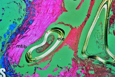
BLOG Nº 13.- .- Foto nº 6.-
Examination with microscopical polarized light to demonstrate the structure of the fibrous thickening sheath (neointima=mfib) around the tip of the lead. (ele= lead). Prof.Garfia.A
Examination with microscopical polarized light to demonstrate the structure of the fibrous thickening sheath (neointima=mfib) around the tip of the lead. (ele= lead). Prof.Garfia.A
 |
| PROF. GARFIA.A BLOG 13 FORENSICPATHOLOGYFORUM |
The fibrous neointima (mfib), surrounding the lead, contains some giant cells (cg) in the proximity to the lead (ele = lead). Prof.Garfia.A