sábado, 28 de febrero de 2009

12.-SUDDEN DEATH AT THE EMERGENCY SERVICE OF AN ANXIOUS PATIENT.Prof.Garfia.A

12.-SUDDEN UNEXPECTED DEATH AT THE HOSPITAL EMERGENCY SERVICE OF AN WOMAN TREATED FOR ANXIETY NEUROSIS:HISTOPATHOLOGICAL FINDINGS.
Prof.Garfia.A


     Patients who suffer serious psychiatric problems, and those others whose symptoms are half way between purely emotional disorders and those dependent on somatization of psychological conflicts, make up a group of patients for whom diagnosis requires special attention and effort. The first form a group at high risk of unexpected death from illnesses so common that it is really surprising that they are not diagnosed and treated in time. The second are faced with the difficulty at diagnosis of differentiating between the truly emotional and the somatic, with the secondary danger of obviating the real somatic cause of some clinical state, with symptoms easily labelled as psychological. At times reality asserts itself drastically, and is legally conflictive for the doctor or the hospital team. 
    It is   presented a case of unexpected death, which took place at the Medical Emergency Service, of a patient diagnosed with anxiety neurosis. 
  The forensic autopsy discovered the existence of a pheochromocytoma of the left adrenal gland .
      Cases of sudden unexpected death associated with the tumor pathology occur seldom. Generally, tumors can causes "truly" sudden death when they provocated serious circulatories problems, such as brain haemorrhages -leucemies- or fatal haemoptisis; massive pulmonary embolization and/or tumoral pulmonary embolization ( liver and digestive system tumors); or massive carcinomatosis ( primary tumors found in the breast, digestive system, pancreas and genital organs).This tumor, the pheochromocytoma, can produce serious cardiovascular pathology, inclusive sudden death, due to the excessive producction of chemical messengers which can produce an overstimulation in the Cardiovascular System, originating a toxic nor-epinephrin myocarditis - due to a direct toxic effect on the myocardiocites; between these messengers are the Adrenalin and Nor-adrenalin and the peptide named adrenomedulin.

CASE REPORT

   In this report it is presented a case of a woman, 50 years old, who was attended at the Hospital Emergency Service at 3.00 hours in the morning. She had a clinical syntomatology consisted of: abdominal pain, dyspnea, and vomiting with blood- tinged froth. The arterial pression was 12/6 and the ECG presented sinusal rythm 120 x. The woman go to the observation room and presented a little later, progressive dyspnea and the followings parameters: Ph 7.21; PCO2 33; PO2 50; HCO3- 13; and O2 saturation 77%. Posteriorly,the woman presented two episodes of cardiac arrest, being necessary aplication of avanced RCP. The patient was transported to the Unit Care and connected to assisted ventilation; in that moment, the arterial pression was 60/0, the ECG shows a sinusal rythms to 130x', a Glasgow of 3 and arreactive mydriasis. Two hours later the patient was pronounced dead. A clinical diagnosis of cardiogenic shock and pulmonary edema of cardiogenic origin was emitted.The family solicited a legal autopsy.
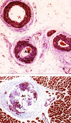
     The macroscopic examination demonstrated the existence of a tumor located in the left renal fossa of 8x5x4,5 cm of maximal diameters (Foto nº 1). Both kidneys were normal.The lungs showed pulmonary edema, and the rest of the organs were normal. Microscopically the epicardial and intramural arterioles in the heart showed hypertensive changes (see Foto nº 2) and also the renal arterioles. The tumor was rounded for a capsule of conective tissue but we can found some tumoral cells penetrating the capsule.Tumor cells were polygonals and they had an ample cytoplasm, brownish, occupied by acidophile granulations.The nuclei were rounded and sometimes giant and pleomorphic. Inside the tumor we found some ganglionar cells and also nests of the glomerulous cells of the adrenal gland ( see Fotos 4-5). The heart shows very abundant focus of myocardiocites presenting contraction band necrosis which do not show a coronary distribution pattern dependence. A diagnostic of epinephrine myocarditis (focal myocarditis) was emitted and was considered the cause of death.


PROF. GARFIA.A
BLOG 12
FORENSICPATHOLOGYFORUM
FIG. 1 .-Macroscopical aspect of the pheochromocytoma (feo), located in the left renal fossa. Left kidney (ri).Prof. Garfia.A


PROF. GARFIA.A
BLOG 13
FORENSICPATHOLOGYFORUM
FIG.2.- Epicardial (above) and intramural (below), arterioles showing hypertensive changes.Prof.Garfia.A


PROF. GARFIA.A
BLOG 13
FORENSICPATHOLOGYFORUM
FIG.-3 Multifocal contracction band necrosis of myocardiocytes (arrows) expression of a ephinefrine myocarditis.
Prof.Garfia.A


PROF. GARFIA.A
BLOG 13
FORENSICPATHOLOGYFORUM
 FIG.4 .- Showing nest of tumoral cells with poligonal form. Tumoral capsule (cap).Prof.Garfia.A


PROF. GARFIA.A
BLOG 13
FORENSICPATHOLOGYFORUM

Fig.5.- Nest of tumoral and ganglionar cells inside the tumor. Arrows showing rests of glomerulous cells of the adrenal gland. Prof.Garfia.A


REFERENCES

1. Garfia A, Borondo J.- Muerte súbita en Patología Forense. Rev Esp Med Legal.Enero-Junio 1989; pp. 31-43.

2. Garfia A.- Glomus tissue in the vicinity of the human carotid sinus. Journal of Anatomy 130: 1-12 (1980).

3.- Garfia A.- En Böck, P.- The Paraganglia. Handbuch der mikroskopischen Anatomie. Band 6. Blutgefäß- und Lymphgefäßapparat. 8 Teil.Springer-Verlag. Berlin1982. pp.89.

4.- Garfia A.- Muerte Súbita o Retardada asociada a la Patología del Compartimento Lateral del cuello: el papel fisiopatológico de los Barorreceptores del Seno Carotídeo y de los Quimiorreceprores del Glomus Caroticum. Cuad Med For 16-17. 1999. 85-153

5.-Garfia et al.-Vascular lesions in the intestinal ischaemia induced by Cocaine-Alcohol Abuse:Report of a Fatal Case due to Overdose.Journal of Forensic Science.Vol 35.nº 3.1990

6. Henle J.- Allgemeine Anatomie. Leipzig 1841.

7. Henle J.- Über die Gewebe der Nebenniere und Hypophyse. Z rat Med 24, 143-152 (1865).

8. Rosai J.- Ackerman´s Surgical Pathology. 7ª Ed. Ed Mosby C. Washington D.C. 1989.

9. Cotran R, Kumar V, Robbins S.- Pathological Basis of Disease. Interamericana. 1995.

10. Silver MD, Gotlieb AI, Schoen FJ.- Cardiovascular Pathology. Churchill Livingstone.N.Y. 2001.

11. Karch SB .- The Pathology of Drug Abuse. 3ª Ed. CRC Press. 2002. pp. 104.

12. Szakacs, J and Cannon, A.- L-Norepinephrine myocarditis. Am J Clin Pathol. 30, pp. 425-434.

13. Van Vliet, Burchell HB, Titus JL.- Focal myocarditis associated with pheochromocytoma. N Engl J Med 274:1102-1105. 1966.

14. Jessurun CR, Adam K, Moisek J Jr, Wilansky S.- Pheochromocytoma-induced myocardial infarction in pregnancy. Tex Heart Inst J 20:120-124;1993.

15. Morris K, McDevitt B.- Pheochromocytoma presenting as a case of mesenteric vascular occlusion. Ir Med J 78:356-358.1985.

16. Gulliford MC, Hawkins CP, Murphy RP.- Spontaneous dissection of the carotid artery and pheochromocytoma. Br J HospMed 35: 416-420, 1986.

17. DeSouza TG, Berlad L, Shaphiro K et al.,- Pheochromocytoma and multiple intracerebral aneurysms. J Pediatr 108: 947-950. 1986.

viernes, 27 de febrero de 2009

11.-SUDDEN DEATH IN ADOLESCENT.PROF.GARFIA.A

11.-SUDDEN AND UNEXPECTED DEATH IN AN ADOLESCENT:INTESTINAL DUPLICATION. Prof.GARFIA.A

CASE REPORT
     It is presented a case of sudden unexpected death, in a 19 year-old adolescent, who in the hours prior to his death presented a clinical picture of dizziness, nausea and vomiting which was treated at the First-Aid Centre with analgesics and antispasmodics. After a few hours the patient was again taken to the Medical Centre by his relatives, by then unconscious. In spite of resuscitation measures the patient presented cardio-respiratory arrest and sudden death. During the autopsy the existence of a mesenteric tumor was discovered in the distal ileum. The corpse gave off a peculiar smell which reminded the Prosector of the typical smell of some organophosphorous pesticides. The following diagnoses for the cause of death were put forward in the autopsy report:

1.- Septic shock due to intestinal abscess. 
2.-Mortal poisoning by pesticides. 
3.-Sudden death of cardiac origin. 

   The chemical investigation resulted negative. The histopathologic study did not show cardiac lesions which could justify the death. The investigation of the intestinal tumor showed that it was a cystic intestinal duplication, with a slight chronic inflamation of the intestinal wall duplication. Perforations, haemorrhage, ischaemic or vascular intestinal lesions which could have justified sudden unexpected death -with digestive causes- were not seen.
The Cause and Manner of Death were considered undetermined.

PROF. GARFIA.A
BLOG 11
FORENSICPATHOLOGYFORUM
FIG. 1.- To show the macroscopical aspect of the cystic intestinal duplication. The cystic wall was similar to the intestinal wall and we found -inside the cyst- a sebaous-gelatinous material brownish. 
Prof.Garfia.A


PROF. GARFIA.A
BLOG 11
FORENSICPATHOLOGYFORUM
FIG.2 .-To show the intestinal normal wall (above) and the cystic intestinal duplication wall (down).Prof. Garfia.A
LI.- Intestinal normal lumen.
LDU.- Lumen of the cystic duplication.
M.- Mucosae.
S.-Submucosae.
MI.-Internal Muscularis.
ME.- External Muscularis which is shared between the normal intestinal wall and the cystic wall duplication.


PROF. GARFIA.A
BLOG 11
FORENSICPATHOLOGYFORUM


FIG.3.- The cystic mucosae( m) appeared constituted by several layers of macrophagical cells (arrows) and cellular necrotic debris on the luminal surface. Prof.Garfia.A


PROF. GARFIA.A
BLOG 11
FORENSICPATHOLOGYFORUM


FIG. 4.- Shows the external cystic wall. Mi= internal muscularis.ME=external muscularis. SP= peritoneal serous. Note the slight linfocitary infiltration in the muscularis wall and focally in the peritoneal serous. Prof.Garfia.A

martes, 24 de febrero de 2009

10.-FATAL AORTIC STAB WOUND.Prof.Garfia.A

10.-FATAL AORTIC STAB WOUND.
Prof.Garfia.A

CASE REPORT

A man 27 years old, who was a drug abuse addict, was stabbed -on the street during an arguement with a man part of a group- with a self-made weapon consisted of a wooden bar, 1.5 mtrs long which was supported, at one end with a pointed knife and the other end with a sharp hammer edge. The homicide was arrested and the weapon found at his house. 
     The autopsy demonstrate an incise wound in the right side of the thorax which penetrated the right lung and the right heart ventricle. The pericardiac sac shows a haematoma, at tension, and at the aortic wall a dissecting haematoma. We received the heart with the dissection made following the haemodinamic outflow with exposition and opening of the aortic valve.    Macroscopically, the heart -260 g - showed a stab wound in the anterior wall of the right ventricle, near the atrioventricular groove, which penetrated into the aorta where could be seen an entrance wound, and an exit wound located at the posterior wall of the aorta ( see. fig.2); itNegrita measured 1 cm long and orientated parallel to the aortic cusps of the aortic valve which penetrated through the aortic wall (trans-aortic stab wound); the exit wound in the posterior side of the aortic wall was plugged with the dissecting haematome.


PROF. GARFIA.A
BLOG 11
FORENSICPATHOLOGYFORUM
Foto 1,2,3.- Weapon used for the murdered, self-made, with a hammer in one extreme and a knife in the other. The bar was 1.5 mtrs long.
Prof. Garfia.A


PROF. GARFIA.A
BLOG 11
FORENSICPATHOLOGYFORUM

Foto 4.-Stab aortic wound. Note the anterior face of the exit wound in the posterior aortic wall. The exit wound is plugged due to the dissecting haematome.(asterisc). 
Prof.Garfia.A

9.- ADENITIS TUBERCULOSAS PERIAÓRTICAS EN SUJETO DROGADICTO.PROF.GARFIA.A

9.-ADENOMEGALIAS PERIAÓRTICAS EN SUJETO DROGADICTO VIH +.
Prof. Garfia.A

CASE REPORT

    Varón de 30 años, adicto a la heroína, VIH +, que en el momento del fallecimiento se encontraba en tratamiento con metadona. 
     En la necropsia se demostraron adenomegalias, de más de tres centímetros y de consistencia aumentada, que afectaban a las cadenas ganglionares preaórticas, aórtico-laterales y retroaórticas, del grupo linfático lumbar. Macroscópicamente, la aorta abdominal y segmentos de la vena cava inferior, aparecían rodeados por un manguito perivascular, segmentario, adenomegálico, que constituía una auténtica envoltura tubular periaórtica. El estudio microscópico puso de manifiesto la existencia de una linfadenitis granulomatosa caseificante; alrededor de los extensos focos de necrosis existía un infiltrado inflamatorio constituído por linfocitos y células plasmáticas, así como por células gigantes multinucleadas, escasas. La tinción, con la técnica de Ziehl-Neelsen demostró la existencia de abundantes bacilos localizados en el citoplasma de células de características macrofágicas. 
      En los sujetos vivos, la punción aspirativa ganglionar, mediante aguja fina, ha permitido definir en el Sida un patrón denominado linfadenitis necrosante, que se caracteriza, en el examen citológico del material obtenido por punción, por la presencia de grandes cantidades de material necrótico -sobre una base granular difusa de color rosa a púrpura- con la tinción de May-Grünwald-Giemsa. El hallazgo de una linfadenitis necrosante es suficiente, en clínica, para iniciar un tratamiento con tuberculostáticos en los pacientes infectados por el VIH. Si no fuese posible la realización de la punción aspirativa, o de la biopsia, el diagnóstico diferencial con otras causas de adenomegalias, tales como: toxoplasmosis, sífilis, leishmaniasis, síndrome de Epstein-Barr, etc. se deberá realizar mediante estudio serológico excluyente, por el PPD (Mantoux) y por la búsqueda del bacilo de Koch en otros órganos (pulmón, por la asociación tan frecuente de la tuberculosis ganglionar con la pulmonar) mediante estudio en aspirado gástrico, esputo, hemocultivo y orina. 
     En los enfermos de SIDA el PPD suele ser negativo a partir del estadio IV, independientemente de que exista tuberculosis activa. Por otro lado, la falta de cooperación entre los linfocitos T y B (secundaria a la afectación de los linfocitos CD4 por el VIH) impide obtener respuestas IgM de fase aguda en las seroaglutinaciones, por lo que debe tenerse en cuenta que una serología negativa no excluye una enfermedad, en el contexto del SIDA. 
     El Mycobacterium avium y el Mycobacterium intracellulare son dos micobacterias muy relacionadas entre sí, que se han agrupado y se conocen con el nombre común de Mycobacterium avium complex (MAC o MAI). El MAI es la micobacteria más frecuentemente encontrada en los enfermos de SIDA y la causa más frecuente de infección bacteriana diseminada dentro de este grupo. 
     La forma diseminada de linfadenitis por MAI suele ocurrir en los estadios terminales de esta enfermedad. El MAI es una micobacteria que se encuentra en el suelo, en el agua y en las plantas y es transportada por los pájaros y los animales de las granjas. La infección humana por MAI es poco frecuente y se consideran no patógenos para el hombre u ocasionalmente oportunistas. En los pacientes inmunodeprimidos, no SIDA dependientes, la infección por MAI puede originar una respuesta granulomatosa caseificante aunque esta respuesta no es frecuente en los enfermos de SIDA, tal como sucede en este caso. El hallazgo morfológico diagnóstico es la presencia de bacilos teñidos de rojo con la técnica de Ziehl-Neelsen localizados en el interior del citoplasma de células macrofágicas. Los bacilos se tiñen , también, con la técnica de Giemsa, con el PAS, con la plata-metenamina, y con la tinción de Brown-Hoops (Hum Pathol 18: 709-714,1987. y Am J Clin Pathol 85: 233-235, 1986).


PROF. GARFIA.A
BLOG 9
FORENSICPATHOLOGYFORUM
FOTO A


PROF. GARFIA.A
BLOG 9
FORENSICPATHOLOGYFORUM
FOTO B
FOTOS A Y B.- CORRESPONDEN A CORTES TRANSVERSALES (A) Y LONGITUDINALES (B) DE LA AORTA ABDOMINAL.NÓTESE EL MANGUITO PERIAÓRTICO CONSTITUIDO POR LAS NUMEROSAS ADENOMEGALIAS (GL), DE LAS CADENAS PERIAÓRTICAS LUMBARES. Prof.GARFIA.A


PROF. GARFIA.A
BLOG 11
FORENSICPATHOLOGYFORUM

FOTO C.- Corte microscópico que muestra la adenitis granulomatosa necrotizante. Las adenomegalias aparecen constituidas por folículos linfoides hiperplásicos que presentan necrosis central (flechas), rodeada por una corona linfocitaria, mal conformada,en el seno de la cual se detectan células gigantes multinucleadas, de Langhans. Tricrómico 150x. Prof.GARFIA.A


PROF. GARFIA.A
BLOG 11
FORENSICPATHOLOGYFORUM

FOTO D.- Detalle de dos células gigantes multinucleadas. Tricrómico 400x.  Prof.Garfia.A


PROF. GARFIA.A
BLOG 11
FORENSICPATHOLOGYFORUM
FOTO E.- Bacilos intracelulares demostrados con la técnica de Ziehl, en las áreas de necrosis, en forma de bastoncillos de color rojo.(flechas).630x.Prof.Garfia.A

viernes, 20 de febrero de 2009

8.-SUDDEN DEATH DUE TO SUBARACHNOID HAEMORRHAGE IN A WOMAN 49 YEARS OLD.Prof.GARFIA.A

8.-POLYCYSTIC KIDNEY DISEASE AND SUDDEN DEATH.
PROF.GARFIA.A

CASE REPORT


Bleeding occurs either into brain substance (brain haemorrhage) or into the subarachnoid space (subarachnoid haemorrhage).The commonest causes of non-traumatic subarachnoid haemorhage (spontaneous haemorrhage) are, in this order:1)rupture of saccular aneurysm (65%) 2) arteriovenous malformation rupture (5%) and 3) extension of intracerebral haematoma usually into ventricles and then into the subarachnoid space ( 5%). About 90% of saccular aneurisms are located in distributions of the internal carotid arteries; the remaining 10% are located on the vertebrobasilar arterial tree.Multiple aneurysms are found in 10-15% of patiens with subarachnoid haemorrhage; these are said to be associated with:
1.- Coarctation of the aorta.
2.- Renal artery stenosis.
3.- Autosomal Dominant Adult Polycystic Kidney Disease ( AD-PCD).
The possible common denominator for the formation of saccular aneurysms is arterial hypertension; however, in AD-PCD there are mutations affecting three genes: PKD1 ( linkage to chromosome 16: 85% of cases); PKD 2 ( 10% of families) and PKD 3. Two of these three genes encode for protein named Policystin I and Policystin II which have an important function in interactions between cells, and between the cells and the extracellular matrix. Abnormal proteins encoded in AD-PCD can play an important role in the origen of saccular aneurysms and in the formation of renal cysts.
In this report it is presented a case of sudden death due to a fatal subarachnoid haemorrage.
The autopsy demonstrates the existence of a AD-PCD and multiple saccular aneurysms in the vertebro-basilar arterial tree -from one of these originated the fatal haemorrhage- and also a Hipertensive Cardiomyopathy (Heart:770g, with biventricular hypertrophy) unknown for the woman and for the family.


FIG.1.- Gross external appearance of the kidneys showing numerous superficial outstanding cysts on the kidneys surface. Prof.Garfia.A

 
FIG.2.-Hemisection of one of the two kidneys shows the different size of the cysts and the colloid content inside some of them ( yellow color as fat aspect).Prof.Garfia.A


FIG. 3 Inside some cysts exists papillar grows (or inversion of the tubular pattern: connective tissue inside, tubular epithelium outside.Arrows.).Prof.Garfia.A


FIG.4.-Renal cysts occupied by proteinaceos fluid showing striking similarities with thyroids colloid. Note the variability of the cysts size and the flattened epithelium lined it. Prof.Garfia.A


FIG.5.-.-Gross external appearance of heart which shows global hypertrophy (weight:770g).
Prof. Garfia.A


FIG.6.-Note the biventricular hypertrophy with severe concentric LV hypertrophy. Prof.Garfia.A


martes, 17 de febrero de 2009

7.-TOXIC EPIDERMAL NECROLYSIS FATAL RELATED TO PHENITOYN ADMINISTRATION.PROF.GARFIA.A

7.-TOXIC EPIDERMAL NECROLYSIS FATAL AND PHENITOYN.
PROF.GARFIA.A

CASE REPORT

     Untowards reactions to drugs are an important medical problem, sometimes followed by a fatal outcome. Although drugs reactions may involve any organ system, cutaneous eruptions drug-induced may result from a drug administered by any route, which reactions can be identified more frequently than other toxics drug-depending effects which occurs in other organs, such as liver and kidney, because their visibility.Toxic epidermal necrolysis (TEN) or Lyell's Syndrome, is one of the most dramatic, the most severe, and one of most often studied cutaneous drug reactions. TEN is characterized by extensive detachment of the epidermis nuder-going full-thickness necrosis.     The drugs more frequently implicated are: anticonvulsivants (phenitoyn, phenobarbital, carbamazepine);antibiotics (ampicillin), sulfonamides and nonsteroids antiinflamatory agents (phenylobutazone, oxyphenbutazone, isoxicam, piroxicam and allopurinol).

We report here a fatal case of TEN related to the anticonvulsivant drug phenitoyn, which was prescribed a man, 74 years old, one month before in order to treat a temporal epilepsy, secondary to a stroke (residual epilepsy).


PROF. GARFIA.A
BLOG 7
FORENSICPATHOLOGYFORUM
FIG.1.-A.-To show a diffuse macular and papular erythematous eruption that rapidly becomes confluent and widespread followed by flaccid fluid filled bullae (B) which rapidly ulcerate, leaving painful raw erosions similar to scalding. Mild pressure on erythematous areas may produce (C) detachment of the epidermis (positive Nikolsky sign) and aspect of wet cloth of the epidermis. As result, there may be large areas of exposed dermis resembling a burn (C). Prof.Garfia.A


PROF. GARFIA.A
BLOG 7
FORENSICPATHOLOGYFORUM

FIG. 2 .- The histopathological features of early lesions of toxic epidermal necrolysis are those of an extreme degree of epidermal keratinocyte necrosis (2A), associated with subepidermal blistering (2B, arrows), as a consecuence of basal cell hydropic degeneration. 
In more mature lesion, there is complete separation of the epidermis (2C) and the roof of the subepidermal bulla is, usually, necrotic. 
2C.-Showing necrotic epidermis completely separated from underlying dermis and to note the paucity of dermal inflamatory infiltrate. Prof.Garfia.A


miércoles, 11 de febrero de 2009

6.-SUDDEN DEATH AND RENAL ONCOCYTOMA.Prof.Garfia.A

6.-ONCOCYTOMA RENAL AND SUDDEN UNEXPECTED DEATH.
PROF.GARFIA.A

CASE REPORT

The case is described of a 57 year-old male who died suddenly in the field where he worked. The body was found several hours later and showed erosion and bruising on the right temple, coinciding with the zone of impact on falling to the ground. From the medical history the family pointed out the existence of a stroke, cardiopathy and non-insulin dependent diabetes.
At autopsy the heart (520 g) shows left concentric ventricular hypertrophy.It was found the existence of a spherical renal tumor about 6 cm in diameter, and orange color on cutting; the centre of the tumor was a cystic cavity lined with whitish fibrous-like tissue.
The probable causal relationships between the existence of the tumor, the hypertensive heart disease and stroke, suffered by the man, and his sudden death is discussed.
Oncocytoma and chromophobe cell carcinoma are two well recognised histological entities in the current classification of renal tumors. Both entities are closely related and arise in the distal portion of the neprhon or in the collector tubules. From a forensic point of view, it is important to point at the incidental autopsy finding of this tumor and, until we know, the classical triad of palpable mass, flank pain or hematuria was absent in this case.
I think that Sudden Death presentation was consecuence of the Hypertensive Heart Disease and the oncocytoma, unfortunatly, an incidental autopsy finding.


BLOG 6
FORENSICPATHOLOGYFORUM
PROF.GARFIA.A
Fig.1.- Gross examination shows a well circumscribed tumor, with encapsulated appearance (arrows), uniforme yellow color and with a central stellate cystic scar (C) which shows striking similarities with a pathological renal pelvis. Is it a kidney (collecting tubules and pelvis) inside the Kidney? Prof.Garfia.A


BLOG 6
FORENSICPATHOLOGYFORUM
PROF. GARFIA.A

Fig. 2.- Detail to show the seudocapsule (arrows), and the central stellate cystic scar (C). Prof.Garfia.A


BLOG 6
FORENSICPATHOLOGYFORUM
PROF. GARFIA.A


Fig.3.-Microscopical aspect of the seudocapsule. (arrows).
Prof.Garfia.A


BLOG 6
FORENSICPATHOLOGYFORUM
PROF. GARFIA.A
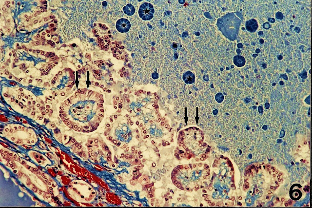
Fig.4.-To show the microscopic tubular pattern inside the tumor (arrow: transversal tubular section from a good differentiated tubule) and differents longitudinal tubular sections (T), where we can see how some tubules are originating from others through a gemmation process (T, at the right side). Haematoxilin-eosin-floxine. Prof.Garfia.A


BLOG 6
FORENSICPATHOLOGYFORUM
PROF. GARFIA.A

Fig. 5.-Reticulin stain to demonstrate basal membranes which show a microscopic tubular pattern delimiting renal tubules (T). Prof.Garfia.A


BLOG 6
FORENSICPATHOLOGYFORUM
PROF. GARFIA.A
 Fig.6.-In others microscopical fields some  structures show striking similarities with primitive glomeruli and contain a tuff of mesangial matrix (M) containing primitive capillaries (at the rihgt figure, just over the letter M. Prof.Garfia.A

5.-TOXIC HAEMORRHAGIC PERIPHERAL NEUROPATHY FOLLOWING POISONING BY RODENTICIDES AND SOLVENTS.Prof. GARFIA.A

5.-TOXIC HAEMORRHAGIC PERIPHERAL NEUROPATHY.
PROF.GARFIA.A

CASE REPORT

It is described a case of mortal poisoning by ingestion of the Rodenticide Brodifacoum, a superwarfarin anticoagulant, together with other drugs, among which were non steroid anti-inflammantory drugs and a domestic organic solvent. The individual died in the Intensive Care Unit, with acute respiratory insufficiency, followed by hepatic and renal failure. At autopsy perineural rounded haemorrhagic lesions were seen, wich affected several tissues, among which were mesenteric fat, the epicardium and the perikidney fat. Microscopy study revealed the existence of a typical acute haemorrhagic lesion, located around the epicardial vegetative nerves and those found in the splanchnic beds. The reason why the peripheral vegetative nerves constitute the target of the toxic effects was attributed to the combined action of the solvents with the superwarfarine drug on the Peripheral Nervous System. It is known that organic solvents produce an acute neuronal lesion, consisting in tumefaction of the axon and the neuronal soma, which leads to the alteration of the system of transport ( neurotubules) along the axon. We believe that -from a physiological point of view - the reactive vascular congestion of the vasa-nervorum - produced as a consecuence of the acute neuronal lesion induced by the solvents, together with the state of hipocoagulability of the blood, due to the ingestion of superwarfarin Brodifacoum, and potentiated by the anticoagulant action on the non steroid anti-inflamatory drugs, make up the intimate mechanism which could explain this characteristic Acute Toxic Haemorrhagic Neuropathy of the peripheral nerves of the Neurovegetative Nervous System. We do not know if the mixed peripheral nerves were affected by this lesion due to absence of samples for a more definitive study. Although most cases of brodifacoum poisoning in humans are non-fatal, this compound, alone, can be deadly because of its very long half-life. 
    In Forensic Pathology we must suspect superwarfarin rodenticides poisoning when confronted with cases of unexplained bleeding. Anticoagulant poisoning can mimic leukemia or infectious diseases such as bacterial sepsis, leptospirosis or rickettsioses; a death scene investigation may provide clues that a person has ingested these substances.

PROF. GARFIA.A
BLOG 5
FORENSICPATHOLOGYFORUM
Fig.1-Showing an Acute Toxic Haemorrhagic Neuropathy affecting a nerve (N) located at the peripancreatic fat. Arrows delimiting the perineurium. Note the aspect " in diana" of the haemorrhage in the plane of this thin slice cut (H= haemorrhagic perineural "sheath" when we thought in three  dimensions). PROF.Garfia.A

sábado, 7 de febrero de 2009

4.-MASSIVE PULMONARY EMBOLIZATION WITH CEREBRAL TISSUE AND LAMELLAR BONE SPICULES DUE TO GUNSHOT WOUND TO THE HEAD.Prof.Garfia.A;Palomo.J.L.

PROF. GARFIA.A
BLOG 4
FORENSICPATHOLOGYFORUM


FIG.-7.-Scheme showing the direction of the bullet in the head: from back to front and from occipital (left) to temporal lobe (right). Prof. Garfia.A


PROF. GARFIA.A
BLOG 7
FORENSICPATHOLOGYFORUM
FIG.-6.- Embolization with cerebral tissue (ETJC) inside a pulmonary artery (AP). Prof.Garfia.A


PROF. GARFIA.A
BLOG 7
FORENSICPATHOLOGYFORUM
FIG.5.-Details to show the brain tissue as a component of the embolus ( arrows: piramidal cortical brain neurons). Klüver-Barrera. Prof.Garfia.A


PROF. GARFIA.A
BLOG 7
FORENSICPATHOLOGYFORUM
FIG.- 3B.- Show the same artery examinated with polarized light (hu lam): the bone spicule is formed of lamellar adult bone. Masson. Prof.Garfia.A

PROF. GARFIA.A
BLOG 7
FORENSICPATHOLOGYFORUM
FIG.3A.-Embolization of bone spicules (arrows) to the lung. Pulmonary artery (stars). Bone spicule (hue- arrows). Masson trichrome stain. Prof.Garfia.A

PROF. GARFIA.A
BLOG 7
FORENSICPATHOLOGYFORUM
Fig. 2.-Liver.Right lobe. Superior face. Penetrating entrance gunshot wound showing stellate aspect. Bullet caliber 22. Prof.Garfia.A


PROF. GARFIA.A
BLOG 7
FORENSICPATHOLOGYFORUM
FIG. 1.- Superior pole of the right kidney. Note the deep niche leaved for the projectil during his trajectory from front to back (arrows). Prof.Garfia.A


Venous and arterial embolism of endogenous tissue components and foreign material must be considered, in forensic pathology, as markers of vital reactions. Pulmonary embolization of cerebral tissue following severe head trauma or due to gunshot wound to the head is uncommonly reported at autopsy. Embolism of bone marrow to the lung is a quite frequent finding after trauma but transport and deposition of solid bone is rarely seen.

CASE REPORT
Prof.Garfia.A
 I report one case of pulmonary embolization with cortical cerebral tissue and with fragments of adult lamellar bone due to gunshot wound to the head in a 32-year-old woman. Brain tissue embolization may have a significant impact on the premortem clinical management of the head trauma patient due to that brain tissue is well known to cause plasma coagulation, shock, and consumptive coagulopathy upon direct contact with the blood stream. These haematologic events have the potential to play a significant role in the morbidity and mortality of head trauma patients. From a statistical and public health perspective, cerebral tissue pulmonary emboli should be sought in all autopsied cases of death due to head injury.