martes, 12 de mayo de 2009

19.-SUICIDE (PARAQUAT- GRAMOXONE) POISONING: CHROMOPATHOLOGICAL FINDINGS.Prof.Garfia.A

19.-SUICIDE PARAQUAT POISONING (GRAMOXONE): CHROMOPATHOLOGICAL ESOPHAGUS-STOMACH IMPREGNATION USEFUL FOR THE DIFFERENTIAL DIAGNOSIS IN CASES OF POISONING DUE TO INGESTION OF CHEMICALS SUBSTANCES.
Prof. Garfia.A


A woman 58, years old, was admitted to the Hospital in a cardiorespiratory arrest after she had drank a glass 250 ml containing Gramoxone ( 1,1´-dipiridil-4,54´dipirilon). A Legal Autopsy was carried out which demonstrated the existence of corrosive lesions around the mouth; the esophageal mucosa was partially desquamated and the gastric mucosa showed one perforation through the posterior wall of the stomach.The more characteristic autopsy finding was the intense green color of the mucosa and submucosa of the oesophagus and stomach wall ; in the case of the stomach, from the mucosa until the serosa, all the gastric wall was coloured in green due to the exit of the herbicide into the abdominal cavity through the perforation of the gastric wall.The Gramoxone pH measured from 6-8 and the gastric content pH was 1. The macroscopic lesions can not be attributed, exclusively, to the ingestion of Gramoxone. Inside the glass from which the woman had drank was found hydrochloric acid that she had used to dissolve the Gramoxon.The colicuative necrosis of the esophageal and gastric mucosa was attributed to the action of the alkaline hydrochloric acid.


FORENSICPATHOLOGYFORUM.BLOGSPOT.COM
BLOG 19
PROF. GARFIA.A
Fig. 1.- This picture was taken from a bottle of Gramoxone Plus which contains Paraquat and Diquat.Prof.Garfia.A


FORENSICPATHOLOGYFORUM.BLOGSPOT.COM
BLOG 19
PROF. GARFIA.A
Fig. 2.- Showing the aspect and the color of the Gramoxone solution. Prof.Garfia.A


FORENSICPATHOLOGYFORUM.BLOGSPOT.COM
BLOG 19
PROF. GARFIA.A
Fig. 3.- Esophagus.Macroscopical aspect showing the mucous membrane, necrotic and detached from the submucous layer - as sphacelous necrotic sheets.The submucous layer and the vascular submucous plexus can be seen under the necrotic sheet. Prof.Garfia.A


FORENSICPATHOLOGYFORUM.BLOGSPOT.COM
BLOG 19
PROF. GARFIA.A
Fig. 4.- Esophagus. Microscopic section showing the transmural necrosis of the esophagus wall. At the left side is located the mucous membrane; at the right side, the submucous and the muscle layer (coloured in red). Note the difficulty of the tissues to take the dyes-that is very typical when a tisular colicuative necrosis occurs - of the Trichromic Stain. Prof.Garfia.A


FORENSICPATHOLOGYFORUM.BLOGSPOT.COM
BLOG 19
PROF. GARFIA.A
Fig. 5.- Esophagus.-Microscopic section of the wall showing the mucous membrane ( on top of the photo), the submucous glands and the severe dilatation of the vessels, due to the caustic effect of the hydrochloric acid. Prof.Garfia. A


FORENSICPATHOLOGYFORUM.BLOGSPOT.COM
BLOG 19
PROF. GARFIA.A

Fig. 6.1- Stomach. Macroscopical aspect. Note the intense green color of the gastric serosa due to the pass of the Gramoxone through the perforation located in the posterior wall of the stomach.Prof.Garfia.A


Fig.6.2.- The gastric mucosa was focally necrotic and coloured in emerald green. In those areas were the mucous membrane layer was absent we can see the submucous vascular net, due to the severe congestion and dilatation of the small submucous vessels. Prof.Garfia.A


FORENSICPATHOLOGYFORUM.BLOGSPOT.COM
BLOG 19
PROF. GARFIA.A
Fig. 7.-Stomach. Showing the microvascular pattern of the submucous layer. The caustic effect of the hydrochloric acid on the microvessels produced vasodilatation and congestion which, together with the coloured effect of the Gramoxone, showed the microvascular pattern of the submucous layer. Normally that is only possible using silver impregnation technics (see: Garfia.A.-Glomus tissue in the vicinity of the human carotid sinus region.Journal of Anatomy. 132:1-12.1980). 
Prof.Garfia.A

sábado, 18 de abril de 2009

18.- LARYNX AS TARGET ORGAN OF ANAPHYLACTIC REACTION IN AN ASTHMATIC PATIENT.Prof.Garfia.A

18.-FATAL OBSTRUCTIVE EDEMA OF THE ARITENOEPIGLOTTIC FOLDS FOLLOWING AN ANAPHYLACTIC REACTION IN A ASMATHIC PATIENT.
PROF.GARFIA.A

Fatal upper airway obstruction can occurs in children and in adults people. In young children is most commonly caused by inhaled foreing bodies, particularly due to food or toy parts. Much less frequently fatal compromise of the superior airway occurs due to an congenital malformation such as a lingual thyrod at the base of the tongue (see:http://www.forensic-histopathology-garfiaa.blogspot.com), from the same author), or a lingual thyroglossal duct cyst. Acquired lesions may also result in fatal airway narrowing and often involve an infectious etiology such as acute epiglottitis, or acute inflamation of a lingual tonsil. Many of these cases occurs in infancy and very early chilhood, although food aspiration, so called "cafe coronary death" may occur at any age. A special chapter in adults it is referred to deaths due to Anaphylactic Reactions after insect bites - wasp, hornets and bees- or deaths following drug administration sometimes difficult to diagnostised due to some drugs are drugs normally utilized againts the anaphylactic process, such as metilprednisolone or other corticoids which utilize "carboximethylcellulose" as disolvent.

CASE REPORT
A finnish man, 58 years old, with pathological antecedents of alergical illnes, arterial hypertension and bronchial asthma presented an undiagnosed clinical picture of pain in the right half of the face, accompanied by important facial and lingual edema. Soon after, the patient went to the Hospital Emergency Service and, after 3 hours, transferred to Intensive Care Unit, where he suffered cardiorespiratory arrest and die.
The medicament treatment guide was the following:

At Home
Urbason....60mg.i.v.
Amoxicillin..875 mg/12 hours.
Antihistamines...(unknown).
Few hours later the clinical picture suffers progresive worsening and the patient go to the Urgency Hospital Service.At the Hospital, the patient presented severe lingual and laringeal edema.The Medical team administrated:
Actocortin...300mg+
Urbason......40 mg, dissolved in a 100ml ampoule of physiological serum. After that, the patient presented a convulsive picture and cardiorespiratory arrest and was sended to the Critic Unit.The orotracheal intubation with a Sheridan tube number 6, was not possible.The patient received the following medication:
Adrenalin: two bolus of 20mg.
Atropine......3mg.
Salbutamol. 3mg
The patient went to cardiovascular arrest and die after 70 min. of cardiopulmonary resuscitation.

FORENSICPATHOLOGYFORUM
BLOG 18
PROF. GARFIA.A


Photo nº 1.-Macroscopical aspect of the larynx, posterior aspect. 
   To note the severe obstructive edema (arrows) of the arytenoepiglottic folds (stars) and the erasure of the cuneiform and corniculate tubercles of the right side due to the brutal suffocating edema, typical of the larynx anaphylactic response. The differential dignosis must be made with the infectious acute fatal epiglottitis, where the affectation of the inflamation affects, more specifically, to the epiglottical structures. Prof.Garfia.A


FORENSICPATHOLOGYFORUM
BLOG 18
PROF. GARFIA.A


PHOTO Nº 2. To show the microscopical aspect of the arytenoepiglottic folds.
Note that the inflamation is due to the edema with scanty cellular componente inside it. This is the second characteristic of the anaphylactic affectation of the larinx (anaphylactic target organ in asthmatic people). Ep=epithelium. Ed= edema.
Prof.Garfia.A


FORENSICPATHOLOGYFORUM
BLOG 18
PROF. GARFIA.A

PHOTO Nº 3.- To show the scanty cellular component (arrows) inside the edema in the anaphylactic reaction. Cells are, principally, leucocytes eosinophiles and plasma cells (the third characteristic of the laringeal anaphilactic reaction). Masson Trichrome-stain.Prof.Garfia.A

jueves, 2 de abril de 2009

17.-FAMILIAL MYOCARDIAL INFARCT.PROF. GARFIA. A

17.-SUDDEN CARDIAC DEATH DUE TO SEVERE CORONARY ATHEROSCLEROSIS IN A YOUNG MAN -26 YEARS OLD.
PROF. GARFIA.A


CASE REPORT


   A 26 year old patient who died as a consequence of myocardial infarction in the presence of witnesses. Two weeks earlier he had visited his family doctor for pain in his shoulder and left arm, which was diagnosed as muscular strain for which the patient was recommended to place his arm in a sling
     The pathological study showed the existence of a myocardial infarct in the interventricular septum and the scar was two weeks old.The left circumflex coronary and the anterior descending arteries, presented severe atheromatous stenosis, of more than 90%. Among the points of interest in the family history, the existence of two episodes of myocardial infarction with hospitalization in the Intensive Care Unit of the victim's elder brother, aged 32 years, due to a myocardial infarct and the death of a first cousin 28 years old - also due to myocardial infarction - were significant. 
     Given the youth of the deceased and his family history, the convenience of carrying out a lipid study was suggested to the family, in view of the suspicion of possible familial hiperlipidaemia.The results of the autopsy, together with the familiy history, the biochemical and the lipid study carried out pointed to the existence of a Combined Primary Familial Hiperlipidaemia. 
    Other possibility to explain the severe atheromatous lesions found in this case would be the existence of an abnormal elevation of Lipoprotein (a) ( > 0.3 g/L), - considered by numerous authors an independent risk factor to have conditioned the apparition of a severe atherosclerotic coronariopathy and sudden cardiac death in young people- was unfortunatelly not determined in this case.


.
FORENSICPATHOLOGYFORUM
BLOG 17
PROF. GARFIA.A


Photo 1.- 26 year old patient. The left anterior descending coronary artery showing severe lumen narrowing due to coronary atherosclerosis (arrow). The red line inside the artery show the residual arterial lumen. Prof.Garfia.A


FORENSICPATHOLOGYFORUM
BLOG 17
PROF. GARFIA.A
 Photo 2.-Microscopical aspect of the same artery showing the severe narrowing of the lumen ( more of 90%) due to the existence of a fibroatheromatous plaque with fibrin deposition (arrows), probably dependent of the organization of a previous trombus (L=lumen). Prof.Garfia.A


FORENSICPATHOLOGYFORUM
BLOG 17
PROF. GARFIA.A
Photo 3.- Microscopical section of the left circumflex coronary artery showing a severe narrowing of the lumen due to the existence of a typical fibrous cap atheroma which contains a necrotic core containing cholesterol clefts, foam cells and fibrous tissue.Prof.Garfia.A


FORENSICPATHOLOGYFORUM
BLOG 17
PROF. GARFIA.A
Photo 4.- Right coronary artery. It shows intimal hyperplasia -with fibrous tissue and foam cells- but the reduction of the arterial lumen (L) is not so severe than in the others coronaries arteries. Focal preatheromatous lesion.
Prof.Garfia.A


FORENSICPATHOLOGYFORUM
BLOG 17
PROF. GARFIA.A
Photo nº.-5.- Showing an intramyocardial arteriole which presents a thicker muscular wall -and penetration of connective tissue from the adventitia- for the age of the patient. Prof.Garfia.A


FORENSICPATHOLOGYFORUM
BLOG 17
PROF. GARFIA.A

Photo nº.6.- This section of the heart shows the localization of the myocardial infarct (arrows) in the interventricular septum.Prof.Garfia.A


FORENSICPATHOLOGYFORUM
BLOG 17
PROF. GARFIA.A

Photo nº. 7.- The mycroscopical study of this area demonstrated the existence of a myocardial infarct -aproximatelly two weeks old- due to the presence of granulation tissue plenty of macrophages cells with cellular debris and neocollagen fibers formation. (IAM= myocardial infarct). 
Prof.Garfia.A

miércoles, 11 de marzo de 2009

15.-CARDIAC ATRIAL TUMOR VERSUS ORGANIZED THROMBUS AND SUDDEN UNEXPECTED DEATH.Prof..Garfia.A

15.-ATRIAL TUMOR AND SUDDEN UNEXPECTED DEATH.
Prof. Garfia.A

     A fithty year old man in good health, suffered an acute heart failure secondary to obstruction of blood flow, and was pronounced dead, at his home. During a legal autopsy a heart weighing 540 g was found, which showed biventricular hypertrophy (RV: 0.8 cm; LV 2.0 cm); the left heart was moderately dilated. 
   At the left atrium was detected a polipoid tumor, 6 cm diameter in its biggest dimension, which was attached, on a broad base, to the atrial septum (fossa ovalis). The tumor had a villous appearance and presented gelatinous areas intercalated with haemorrhagic areas.The left atrium was almost filled by the tumor which extended down into the mitral valve orifice.    The histological appearance of the tumor was very variable in different areas examinated.The bulk of the tumor is made up of a myxoid stroma and embedded in the stroma were the myxoma cells which shows polygonal, spindle or stellate-shaped.The term "lepidic" (scale-like) has been applied to these cells due to their polygonal aspect. In some areas can be seen spaces lined by endothelial cells to which the myxoma cells are loosely attached. Inside the macroscopic haemorrhagic areas the tumor shows numerous vascular spaces of telangiectasis aspect. Also, other elements found in the matrix include haemorrhage foci, old and more recent, located besides the telangiectasis spaces.The differential diagnosis must be made with low-grade sarcomas, so named "myxoid imitators". Sometimes, the distinction between organized thrombus and myxoma, is very difficult. Virtually any cardiac tumor may cause sudden death through a variety of mechanismus including rhythm disturbances, embolization and acute heart failure secondary to obstruction of blood flow. Cardiac myxomas are- generally- tumors found in adults, and present as sudden death in aproximatelly 5% of cases due to embolization to the coronary arteries, acute obstruction of the mitral valve, or also, cerebral embolization
Atrial myxomas are the most common primary benign tumour, show a slight female predominance and are, generally, located in  the left atrium. Generally, myxomas are solitary tumors, however, a familial myxoma syndrome has been described: it is called NAME, (acronyms of N: blue naevi; A: Atrial myxoma; M: mucocutaneous myxoma; E: ephelides) or LAMB ( L= lentigines; A= atrial myxoma; M=mucocutaneous myxomas; B blue naevi). In the familial syndrome the tumours can be multiple and located also in a ventricle.

FORENSICPATHOLOGYFORUM
BLOG 15
PROF. GARFIA.A
Photo nº 1 .-To show the macroscopical aspect of the tumor in the left atrium. Prof.Garfia.A


FORENSICPATHOLOGYFORUM
BLOG 15
PROF. GARFIA.A


Photo nº 2.- Gross Pathology:tumoral surface.
   The tumoral surface - mamelonne- shows different aspects and colours, from the red-wine, and haemorrhagic aspect, until pearly-white colour. Prof.Garfia.A


FORENSICPATHOLOGYFORUM
BLOG 15
PROF. GARFIA.A
Photo nº 3.- Atrial myxoma. 
Shows the hystology of the components of myxoma: free-floating spindle and stellate cells -sometimes syncytial-; myxoid ground substance, and a surface layer.
Prof.Garfia.A


FORENSICPATHOLOGYFORUM
BLOG 15
PROF. GARFIA.A
Photo nº 4.- Shows a very characteristic appearance of myxoma cells arranged in a cuff around a small central space.
Prof.Garfia.A


FORENSICPATHOLOGYFORUM
BLOG 15
PROF. GARFIA.A
 
Photo .- 5.- Atrial myxoma. 
    Some cells show nuclei similar to the Anitschkow cells found in the rheumatic carditis (spindle -shaped cells showing ovoid open vesicular nuclei and condensation of the chromatin toward the nuclear membrane - caterpillar cells). They are considered as a variety of mesenchymall cell readily induced in the connective tissue of the heart, in young individuals, by a wide range of insults. Prof.Garfia.A

martes, 10 de marzo de 2009

14.-TURPENTINE POISONING:REPORT OF A FATAL CASE.Prof.Garfia.A

14.-Fatal Poisoning due to Suicidal Ingestion of Turpentine Solution. Autopsy and Histopathological Findings.
Prof.Garfia.A

A -40 year old- woman was admitted to the Intensive Care Unit with coma and apnea. There was insufficient information about the manner and the speed of coma onset, as she had been found by her relatives, in her room, where she was laying unconcious, on the floor.The woman was under treatment with antidepresive and ansyolitics drugs prescribed by a psychiatrist doctor.The patient was treated with hemoperfusion and was pronounced dead due to multiorganic failure, two hours later. A legal autopsy was done.Turpentine ingestion was suspected from the beginning of the autopsy due to the pine odor of the corpse.The relatives said that they found a bottle of Turpentine beside the patient's bed.
     The more important findings during the autopsy were relative to the gastrointestinal system, specially at the oesophagus and the stomach; both organs show dark blood on the mucosa surface and the stomach was dilated and contained 120 ml of a dark bloody liquid.The organ show several perforations and can be seen gastric contents in the peritoneum with necrosis of the spleen capsule. Samples of gastric fluid and blood contain cyclic terpenos, normal components of pine oil.    Turpentine is a colorless thin transparent oily liquid with a strong specific odor (pine odor; violet odor), insoluble in water but soluble in numerous organic solvents. It is used to dissolve oil-based paints, varnish and grease stains. During acute poisoning with Turpentine, the more important clinical manifestations occur in the gastrointestinal system -nausea, vomiting and diarrhea- and in the Nervous System, as coma or stupor. The acute toxic oral dose has been estimated in excess of 2 mL/kg. Systemic toxicity, when it appears, ocurs two or three hours after the exposure. The main metabolite of monoterpenes (the main constituents in turpentine solutions), named bornylacetate, has a peak excretion in urine the 5th and 6th post-exposure day. Hemoperfusion eliminates turpentine constituents effectively from the blood and only should be applied at an early stage of intoxication before accumulation of the toxins in tissues be established. Inmediate and continous gastric lavage is the most effective therapy.


forensicpathologyforum
blog 14
Prof.Garfia.A


Foto A.- Show a very important gastric dilation due to the colliquative necrosis and gastromalacia of the organ.
Foto B.- Exposed gastric mucosa, after the opening, showing the colliquative necrosis and the gastric hemorrhage. 
Prof.Garfia.A

domingo, 8 de marzo de 2009

13.-AUTOPSY EVALUATION OF INTRACARDIAC DEVICES. I.- PACEMAKERS. Prof.GARFIA.A

BLOG 13.-
INTRACARDIAC DEVICES EVALUATION: A FACE UP TO THE AUTOPSY PATHOLOGIST.
I.- PACEMAKERS.
Prof.GARFIA.A

  Implantable devices for the management of the cardiac illness is increasing; for this reason a pathologist can found an intracardiac device during his professional life. Several types of devices can be found implanted in the heart, such as: cardioverter-defibrillators, pacemakers, prosthetic heart valves, occluder devices, stents, etc. The material used for the differents types of devices can be inorganic - polyfluorocarbons, cobalt and titanium, chromium alloys, ceramics- or biologic  (fascia lata, dura, bovine and porcine pericardium,etc).


Pathologist's Role at Autopsy.-
   At the autopsy, the pathologist must examine for degenerative changes in presence of prosthetic valves, and also for ring abcesses, perivalvar leaks, or strut fractures in Björk-Shiley prostheses with occluder escape -which are rare complications of mechanical valves. Degenerative changes with infection and perforation are not unfrequent in bioprosthetic valves. In cases of suspect pacemaker malfunction must be investigated the pulse generator and the leads -it is said: test of the battery, pulse width, sensing function and integrity of leads. Some iatrogenic complications include entrapment of the pacingwire in the tricuspid valve, neointima formation around the lead adjacent to the tricuspide valve and tip, and fibrous thickening at the tip encasing the lead within endocardial tissue (see fotos 5-6-7). 
     In opinion of some authors, these changes are not necessarily associated with the age of the pacemaker and the inclusion of the leads inside the right ventricular wall explains the reason for the problems to extracting pacemakers from living patients.


PROF. GARFIA.A
BLOG 13
FORENSICPATHOLOGYFORUM

BLOG Nº 13.-Foto nº 1.- To show the leads components of a dual chamber pacemaker "in situ", after the opening of the right atrium. Prof.Garfia.A


PROF. GARFIA.A
BLOG 13
FORENSICPATHOLOGYFORUM
BLOG Nº 13.- Foto nº 2.-Sagital section through the right heart in order to follow the course of the pacemaker. AD=right atrium. VD= right ventricle. VDPA= rihgt ventricle anterior wall. VT= mitral valve. Arrows showing insertion points of the leads in the right heart. Prof.Garfia.A


PROF. GARFIA.A
BLOG 13
FORENSICPATHOLOGYFORUM
BLOG Nº 13.-  Foto nº 3.- 
Detail to show the organic -fibrin and platelets- sheath created around the metallic lead which lies in the atrium in this dual chamber pacemaker. Prof. Garfia.A


PROF. GARFIA.A
BLOG 13
FORENSICPATHOLOGYFORUM
BLOG Nº 13.- Foto nº4.-
Detail of the friable sheath around the metallic envelope of the atrial lead. Prof.Garfia.A 


PROF. GARFIA.A
BLOG 13
FORENSICPATHOLOGYFORUM

BLOG Nº 13.-Foto nº 5.- To demonstrates the ventricular lead, which has incited a fibrotic reaction (arrow) in the right ventricular wall (neointima formation). This fibrous reaction may make extraction of the device difficult - must be necessary open heart surgery to do it. Prof. Garfia.A


PROF. GARFIA.A
BLOG 13
FORENSICPATHOLOGYFORUM

BLOG Nº 13.- .- Foto nº 6.-
Examination with microscopical polarized light to demonstrate the structure of the fibrous thickening sheath (neointima=mfib) around the tip of the lead. (ele= lead). Prof.Garfia.A


PROF. GARFIA.A
BLOG 13
FORENSICPATHOLOGYFORUM
BLOG Nº 13.-  Foto nº 7.- 
The fibrous neointima (mfib), surrounding the lead, contains some giant cells (cg) in the proximity to the lead (ele = lead). Prof.Garfia.A

sábado, 28 de febrero de 2009

12.-SUDDEN DEATH AT THE EMERGENCY SERVICE OF AN ANXIOUS PATIENT.Prof.Garfia.A

12.-SUDDEN UNEXPECTED DEATH AT THE HOSPITAL EMERGENCY SERVICE OF AN WOMAN TREATED FOR ANXIETY NEUROSIS:HISTOPATHOLOGICAL FINDINGS.
Prof.Garfia.A


     Patients who suffer serious psychiatric problems, and those others whose symptoms are half way between purely emotional disorders and those dependent on somatization of psychological conflicts, make up a group of patients for whom diagnosis requires special attention and effort. The first form a group at high risk of unexpected death from illnesses so common that it is really surprising that they are not diagnosed and treated in time. The second are faced with the difficulty at diagnosis of differentiating between the truly emotional and the somatic, with the secondary danger of obviating the real somatic cause of some clinical state, with symptoms easily labelled as psychological. At times reality asserts itself drastically, and is legally conflictive for the doctor or the hospital team. 
    It is   presented a case of unexpected death, which took place at the Medical Emergency Service, of a patient diagnosed with anxiety neurosis. 
  The forensic autopsy discovered the existence of a pheochromocytoma of the left adrenal gland .
      Cases of sudden unexpected death associated with the tumor pathology occur seldom. Generally, tumors can causes "truly" sudden death when they provocated serious circulatories problems, such as brain haemorrhages -leucemies- or fatal haemoptisis; massive pulmonary embolization and/or tumoral pulmonary embolization ( liver and digestive system tumors); or massive carcinomatosis ( primary tumors found in the breast, digestive system, pancreas and genital organs).This tumor, the pheochromocytoma, can produce serious cardiovascular pathology, inclusive sudden death, due to the excessive producction of chemical messengers which can produce an overstimulation in the Cardiovascular System, originating a toxic nor-epinephrin myocarditis - due to a direct toxic effect on the myocardiocites; between these messengers are the Adrenalin and Nor-adrenalin and the peptide named adrenomedulin.

CASE REPORT

   In this report it is presented a case of a woman, 50 years old, who was attended at the Hospital Emergency Service at 3.00 hours in the morning. She had a clinical syntomatology consisted of: abdominal pain, dyspnea, and vomiting with blood- tinged froth. The arterial pression was 12/6 and the ECG presented sinusal rythm 120 x. The woman go to the observation room and presented a little later, progressive dyspnea and the followings parameters: Ph 7.21; PCO2 33; PO2 50; HCO3- 13; and O2 saturation 77%. Posteriorly,the woman presented two episodes of cardiac arrest, being necessary aplication of avanced RCP. The patient was transported to the Unit Care and connected to assisted ventilation; in that moment, the arterial pression was 60/0, the ECG shows a sinusal rythms to 130x', a Glasgow of 3 and arreactive mydriasis. Two hours later the patient was pronounced dead. A clinical diagnosis of cardiogenic shock and pulmonary edema of cardiogenic origin was emitted.The family solicited a legal autopsy.
     The macroscopic examination demonstrated the existence of a tumor located in the left renal fossa of 8x5x4,5 cm of maximal diameters (Foto nº 1). Both kidneys were normal.The lungs showed pulmonary edema, and the rest of the organs were normal. Microscopically the epicardial and intramural arterioles in the heart showed hypertensive changes (see Foto nº 2) and also the renal arterioles. The tumor was rounded for a capsule of conective tissue but we can found some tumoral cells penetrating the capsule.Tumor cells were polygonals and they had an ample cytoplasm, brownish, occupied by acidophile granulations.The nuclei were rounded and sometimes giant and pleomorphic. Inside the tumor we found some ganglionar cells and also nests of the glomerulous cells of the adrenal gland ( see Fotos 4-5). The heart shows very abundant focus of myocardiocites presenting contraction band necrosis which do not show a coronary distribution pattern dependence. A diagnostic of epinephrine myocarditis (focal myocarditis) was emitted and was considered the cause of death.


PROF. GARFIA.A
BLOG 12
FORENSICPATHOLOGYFORUM
FIG. 1 .-Macroscopical aspect of the pheochromocytoma (feo), located in the left renal fossa. Left kidney (ri).Prof. Garfia.A


PROF. GARFIA.A
BLOG 13
FORENSICPATHOLOGYFORUM
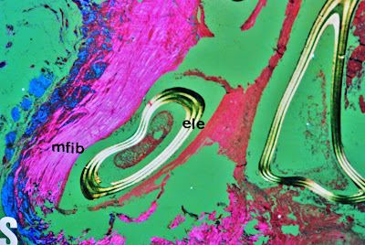
FIG.2.- Epicardial (above) and intramural (below), arterioles showing hypertensive changes.Prof.Garfia.A


PROF. GARFIA.A
BLOG 13
FORENSICPATHOLOGYFORUM
FIG.-3 Multifocal contracction band necrosis of myocardiocytes (arrows) expression of a ephinefrine myocarditis.
Prof.Garfia.A


PROF. GARFIA.A
BLOG 13
FORENSICPATHOLOGYFORUM
 FIG.4 .- Showing nest of tumoral cells with poligonal form. Tumoral capsule (cap).Prof.Garfia.A


PROF. GARFIA.A
BLOG 13
FORENSICPATHOLOGYFORUM

Fig.5.- Nest of tumoral and ganglionar cells inside the tumor. Arrows showing rests of glomerulous cells of the adrenal gland. Prof.Garfia.A


REFERENCES

1. Garfia A, Borondo J.- Muerte súbita en Patología Forense. Rev Esp Med Legal.Enero-Junio 1989; pp. 31-43.

2. Garfia A.- Glomus tissue in the vicinity of the human carotid sinus. Journal of Anatomy 130: 1-12 (1980).

3.- Garfia A.- En Böck, P.- The Paraganglia. Handbuch der mikroskopischen Anatomie. Band 6. Blutgefäß- und Lymphgefäßapparat. 8 Teil.Springer-Verlag. Berlin1982. pp.89.

4.- Garfia A.- Muerte Súbita o Retardada asociada a la Patología del Compartimento Lateral del cuello: el papel fisiopatológico de los Barorreceptores del Seno Carotídeo y de los Quimiorreceprores del Glomus Caroticum. Cuad Med For 16-17. 1999. 85-153

5.-Garfia et al.-Vascular lesions in the intestinal ischaemia induced by Cocaine-Alcohol Abuse:Report of a Fatal Case due to Overdose.Journal of Forensic Science.Vol 35.nº 3.1990

6. Henle J.- Allgemeine Anatomie. Leipzig 1841.

7. Henle J.- Über die Gewebe der Nebenniere und Hypophyse. Z rat Med 24, 143-152 (1865).

8. Rosai J.- Ackerman´s Surgical Pathology. 7ª Ed. Ed Mosby C. Washington D.C. 1989.

9. Cotran R, Kumar V, Robbins S.- Pathological Basis of Disease. Interamericana. 1995.

10. Silver MD, Gotlieb AI, Schoen FJ.- Cardiovascular Pathology. Churchill Livingstone.N.Y. 2001.

11. Karch SB .- The Pathology of Drug Abuse. 3ª Ed. CRC Press. 2002. pp. 104.

12. Szakacs, J and Cannon, A.- L-Norepinephrine myocarditis. Am J Clin Pathol. 30, pp. 425-434.

13. Van Vliet, Burchell HB, Titus JL.- Focal myocarditis associated with pheochromocytoma. N Engl J Med 274:1102-1105. 1966.

14. Jessurun CR, Adam K, Moisek J Jr, Wilansky S.- Pheochromocytoma-induced myocardial infarction in pregnancy. Tex Heart Inst J 20:120-124;1993.

15. Morris K, McDevitt B.- Pheochromocytoma presenting as a case of mesenteric vascular occlusion. Ir Med J 78:356-358.1985.

16. Gulliford MC, Hawkins CP, Murphy RP.- Spontaneous dissection of the carotid artery and pheochromocytoma. Br J HospMed 35: 416-420, 1986.

17. DeSouza TG, Berlad L, Shaphiro K et al.,- Pheochromocytoma and multiple intracerebral aneurysms. J Pediatr 108: 947-950. 1986.

viernes, 27 de febrero de 2009

11.-SUDDEN DEATH IN ADOLESCENT.PROF.GARFIA.A

11.-SUDDEN AND UNEXPECTED DEATH IN AN ADOLESCENT:INTESTINAL DUPLICATION. Prof.GARFIA.A

CASE REPORT
     It is presented a case of sudden unexpected death, in a 19 year-old adolescent, who in the hours prior to his death presented a clinical picture of dizziness, nausea and vomiting which was treated at the First-Aid Centre with analgesics and antispasmodics. After a few hours the patient was again taken to the Medical Centre by his relatives, by then unconscious. In spite of resuscitation measures the patient presented cardio-respiratory arrest and sudden death. During the autopsy the existence of a mesenteric tumor was discovered in the distal ileum. The corpse gave off a peculiar smell which reminded the Prosector of the typical smell of some organophosphorous pesticides. The following diagnoses for the cause of death were put forward in the autopsy report:

1.- Septic shock due to intestinal abscess. 
2.-Mortal poisoning by pesticides. 
3.-Sudden death of cardiac origin. 

   The chemical investigation resulted negative. The histopathologic study did not show cardiac lesions which could justify the death. The investigation of the intestinal tumor showed that it was a cystic intestinal duplication, with a slight chronic inflamation of the intestinal wall duplication. Perforations, haemorrhage, ischaemic or vascular intestinal lesions which could have justified sudden unexpected death -with digestive causes- were not seen.
The Cause and Manner of Death were considered undetermined.

PROF. GARFIA.A
BLOG 11
FORENSICPATHOLOGYFORUM
FIG. 1.- To show the macroscopical aspect of the cystic intestinal duplication. The cystic wall was similar to the intestinal wall and we found -inside the cyst- a sebaous-gelatinous material brownish. 
Prof.Garfia.A


PROF. GARFIA.A
BLOG 11
FORENSICPATHOLOGYFORUM
FIG.2 .-To show the intestinal normal wall (above) and the cystic intestinal duplication wall (down).Prof. Garfia.A
LI.- Intestinal normal lumen.
LDU.- Lumen of the cystic duplication.
M.- Mucosae.
S.-Submucosae.
MI.-Internal Muscularis.
ME.- External Muscularis which is shared between the normal intestinal wall and the cystic wall duplication.


PROF. GARFIA.A
BLOG 11
FORENSICPATHOLOGYFORUM


FIG.3.- The cystic mucosae( m) appeared constituted by several layers of macrophagical cells (arrows) and cellular necrotic debris on the luminal surface. Prof.Garfia.A


PROF. GARFIA.A
BLOG 11
FORENSICPATHOLOGYFORUM


FIG. 4.- Shows the external cystic wall. Mi= internal muscularis.ME=external muscularis. SP= peritoneal serous. Note the slight linfocitary infiltration in the muscularis wall and focally in the peritoneal serous. Prof.Garfia.A

martes, 24 de febrero de 2009

10.-FATAL AORTIC STAB WOUND.Prof.Garfia.A

10.-FATAL AORTIC STAB WOUND.
Prof.Garfia.A

CASE REPORT

A man 27 years old, who was a drug abuse addict, was stabbed -on the street during an arguement with a man part of a group- with a self-made weapon consisted of a wooden bar, 1.5 mtrs long which was supported, at one end with a pointed knife and the other end with a sharp hammer edge. The homicide was arrested and the weapon found at his house. 
     The autopsy demonstrate an incise wound in the right side of the thorax which penetrated the right lung and the right heart ventricle. The pericardiac sac shows a haematoma, at tension, and at the aortic wall a dissecting haematoma. We received the heart with the dissection made following the haemodinamic outflow with exposition and opening of the aortic valve.    Macroscopically, the heart -260 g - showed a stab wound in the anterior wall of the right ventricle, near the atrioventricular groove, which penetrated into the aorta where could be seen an entrance wound, and an exit wound located at the posterior wall of the aorta ( see. fig.2); itNegrita measured 1 cm long and orientated parallel to the aortic cusps of the aortic valve which penetrated through the aortic wall (trans-aortic stab wound); the exit wound in the posterior side of the aortic wall was plugged with the dissecting haematome.


PROF. GARFIA.A
BLOG 11
FORENSICPATHOLOGYFORUM
Foto 1,2,3.- Weapon used for the murdered, self-made, with a hammer in one extreme and a knife in the other. The bar was 1.5 mtrs long.
Prof. Garfia.A


PROF. GARFIA.A
BLOG 11
FORENSICPATHOLOGYFORUM

Foto 4.-Stab aortic wound. Note the anterior face of the exit wound in the posterior aortic wall. The exit wound is plugged due to the dissecting haematome.(asterisc). 
Prof.Garfia.A