 |
| PROF. GARFIA.A BLOG 4 FORENSICPATHOLOGYFORUM |
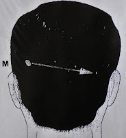
FIG.-7.-Scheme showing the direction of the bullet in the head: from back to front and from occipital (left) to temporal lobe (right). Prof. Garfia.A
 |
| PROF. GARFIA.A BLOG 7 FORENSICPATHOLOGYFORUM |
 |
| PROF. GARFIA.A BLOG 7 FORENSICPATHOLOGYFORUM |
 |
| PROF. GARFIA.A BLOG 7 FORENSICPATHOLOGYFORUM |
 |
| PROF. GARFIA.A BLOG 7 FORENSICPATHOLOGYFORUM |
 |
| PROF. GARFIA.A BLOG 7 FORENSICPATHOLOGYFORUM |
 |
| PROF. GARFIA.A BLOG 7 FORENSICPATHOLOGYFORUM |
Venous and arterial embolism of endogenous tissue components and foreign material must be considered, in forensic pathology, as markers of vital reactions. Pulmonary embolization of cerebral tissue following severe head trauma or due to gunshot wound to the head is uncommonly reported at autopsy. Embolism of bone marrow to the lung is a quite frequent finding after trauma but transport and deposition of solid bone is rarely seen.
CASE REPORT
Prof.Garfia.A
Prof.Garfia.A
I report one case of pulmonary embolization with cortical cerebral tissue and with fragments of adult lamellar bone due to gunshot wound to the head in a 32-year-old woman. Brain tissue embolization may have a significant impact on the premortem clinical management of the head trauma patient due to that brain tissue is well known to cause plasma coagulation, shock, and consumptive coagulopathy upon direct contact with the blood stream. These haematologic events have the potential to play a significant role in the morbidity and mortality of head trauma patients. From a statistical and public health perspective, cerebral tissue pulmonary emboli should be sought in all autopsied cases of death due to head injury.


