miércoles, 11 de marzo de 2009

15.-CARDIAC ATRIAL TUMOR VERSUS ORGANIZED THROMBUS AND SUDDEN UNEXPECTED DEATH.Prof..Garfia.A

15.-ATRIAL TUMOR AND SUDDEN UNEXPECTED DEATH.
Prof. Garfia.A

     A fithty year old man in good health, suffered an acute heart failure secondary to obstruction of blood flow, and was pronounced dead, at his home. During a legal autopsy a heart weighing 540 g was found, which showed biventricular hypertrophy (RV: 0.8 cm; LV 2.0 cm); the left heart was moderately dilated. 
   At the left atrium was detected a polipoid tumor, 6 cm diameter in its biggest dimension, which was attached, on a broad base, to the atrial septum (fossa ovalis). The tumor had a villous appearance and presented gelatinous areas intercalated with haemorrhagic areas.The left atrium was almost filled by the tumor which extended down into the mitral valve orifice.    The histological appearance of the tumor was very variable in different areas examinated.The bulk of the tumor is made up of a myxoid stroma and embedded in the stroma were the myxoma cells which shows polygonal, spindle or stellate-shaped.The term "lepidic" (scale-like) has been applied to these cells due to their polygonal aspect. In some areas can be seen spaces lined by endothelial cells to which the myxoma cells are loosely attached. Inside the macroscopic haemorrhagic areas the tumor shows numerous vascular spaces of telangiectasis aspect. Also, other elements found in the matrix include haemorrhage foci, old and more recent, located besides the telangiectasis spaces.The differential diagnosis must be made with low-grade sarcomas, so named "myxoid imitators". Sometimes, the distinction between organized thrombus and myxoma, is very difficult. Virtually any cardiac tumor may cause sudden death through a variety of mechanismus including rhythm disturbances, embolization and acute heart failure secondary to obstruction of blood flow. Cardiac myxomas are- generally- tumors found in adults, and present as sudden death in aproximatelly 5% of cases due to embolization to the coronary arteries, acute obstruction of the mitral valve, or also, cerebral embolization
Atrial myxomas are the most common primary benign tumour, show a slight female predominance and are, generally, located in  the left atrium. Generally, myxomas are solitary tumors, however, a familial myxoma syndrome has been described: it is called NAME, (acronyms of N: blue naevi; A: Atrial myxoma; M: mucocutaneous myxoma; E: ephelides) or LAMB ( L= lentigines; A= atrial myxoma; M=mucocutaneous myxomas; B blue naevi). In the familial syndrome the tumours can be multiple and located also in a ventricle.

FORENSICPATHOLOGYFORUM
BLOG 15
PROF. GARFIA.A
Photo nº 1 .-To show the macroscopical aspect of the tumor in the left atrium. Prof.Garfia.A


FORENSICPATHOLOGYFORUM
BLOG 15
PROF. GARFIA.A


Photo nº 2.- Gross Pathology:tumoral surface.
   The tumoral surface - mamelonne- shows different aspects and colours, from the red-wine, and haemorrhagic aspect, until pearly-white colour. Prof.Garfia.A


FORENSICPATHOLOGYFORUM
BLOG 15
PROF. GARFIA.A
Photo nº 3.- Atrial myxoma. 
Shows the hystology of the components of myxoma: free-floating spindle and stellate cells -sometimes syncytial-; myxoid ground substance, and a surface layer.
Prof.Garfia.A


FORENSICPATHOLOGYFORUM
BLOG 15
PROF. GARFIA.A
Photo nº 4.- Shows a very characteristic appearance of myxoma cells arranged in a cuff around a small central space.
Prof.Garfia.A


FORENSICPATHOLOGYFORUM
BLOG 15
PROF. GARFIA.A
 
Photo .- 5.- Atrial myxoma. 
    Some cells show nuclei similar to the Anitschkow cells found in the rheumatic carditis (spindle -shaped cells showing ovoid open vesicular nuclei and condensation of the chromatin toward the nuclear membrane - caterpillar cells). They are considered as a variety of mesenchymall cell readily induced in the connective tissue of the heart, in young individuals, by a wide range of insults. Prof.Garfia.A

martes, 10 de marzo de 2009

14.-TURPENTINE POISONING:REPORT OF A FATAL CASE.Prof.Garfia.A

14.-Fatal Poisoning due to Suicidal Ingestion of Turpentine Solution. Autopsy and Histopathological Findings.
Prof.Garfia.A

A -40 year old- woman was admitted to the Intensive Care Unit with coma and apnea. There was insufficient information about the manner and the speed of coma onset, as she had been found by her relatives, in her room, where she was laying unconcious, on the floor.The woman was under treatment with antidepresive and ansyolitics drugs prescribed by a psychiatrist doctor.The patient was treated with hemoperfusion and was pronounced dead due to multiorganic failure, two hours later. A legal autopsy was done.Turpentine ingestion was suspected from the beginning of the autopsy due to the pine odor of the corpse.The relatives said that they found a bottle of Turpentine beside the patient's bed.
     The more important findings during the autopsy were relative to the gastrointestinal system, specially at the oesophagus and the stomach; both organs show dark blood on the mucosa surface and the stomach was dilated and contained 120 ml of a dark bloody liquid.The organ show several perforations and can be seen gastric contents in the peritoneum with necrosis of the spleen capsule. Samples of gastric fluid and blood contain cyclic terpenos, normal components of pine oil.    Turpentine is a colorless thin transparent oily liquid with a strong specific odor (pine odor; violet odor), insoluble in water but soluble in numerous organic solvents. It is used to dissolve oil-based paints, varnish and grease stains. During acute poisoning with Turpentine, the more important clinical manifestations occur in the gastrointestinal system -nausea, vomiting and diarrhea- and in the Nervous System, as coma or stupor. The acute toxic oral dose has been estimated in excess of 2 mL/kg. Systemic toxicity, when it appears, ocurs two or three hours after the exposure. The main metabolite of monoterpenes (the main constituents in turpentine solutions), named bornylacetate, has a peak excretion in urine the 5th and 6th post-exposure day. Hemoperfusion eliminates turpentine constituents effectively from the blood and only should be applied at an early stage of intoxication before accumulation of the toxins in tissues be established. Inmediate and continous gastric lavage is the most effective therapy.


forensicpathologyforum
blog 14
Prof.Garfia.A


Foto A.- Show a very important gastric dilation due to the colliquative necrosis and gastromalacia of the organ.
Foto B.- Exposed gastric mucosa, after the opening, showing the colliquative necrosis and the gastric hemorrhage. 
Prof.Garfia.A

domingo, 8 de marzo de 2009

13.-AUTOPSY EVALUATION OF INTRACARDIAC DEVICES. I.- PACEMAKERS. Prof.GARFIA.A

BLOG 13.-
INTRACARDIAC DEVICES EVALUATION: A FACE UP TO THE AUTOPSY PATHOLOGIST.
I.- PACEMAKERS.
Prof.GARFIA.A

  Implantable devices for the management of the cardiac illness is increasing; for this reason a pathologist can found an intracardiac device during his professional life. Several types of devices can be found implanted in the heart, such as: cardioverter-defibrillators, pacemakers, prosthetic heart valves, occluder devices, stents, etc. The material used for the differents types of devices can be inorganic - polyfluorocarbons, cobalt and titanium, chromium alloys, ceramics- or biologic  (fascia lata, dura, bovine and porcine pericardium,etc).


Pathologist's Role at Autopsy.-
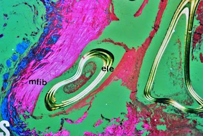
   At the autopsy, the pathologist must examine for degenerative changes in presence of prosthetic valves, and also for ring abcesses, perivalvar leaks, or strut fractures in Björk-Shiley prostheses with occluder escape -which are rare complications of mechanical valves. Degenerative changes with infection and perforation are not unfrequent in bioprosthetic valves. In cases of suspect pacemaker malfunction must be investigated the pulse generator and the leads -it is said: test of the battery, pulse width, sensing function and integrity of leads. Some iatrogenic complications include entrapment of the pacingwire in the tricuspid valve, neointima formation around the lead adjacent to the tricuspide valve and tip, and fibrous thickening at the tip encasing the lead within endocardial tissue (see fotos 5-6-7). 
     In opinion of some authors, these changes are not necessarily associated with the age of the pacemaker and the inclusion of the leads inside the right ventricular wall explains the reason for the problems to extracting pacemakers from living patients.


PROF. GARFIA.A
BLOG 13
FORENSICPATHOLOGYFORUM

BLOG Nº 13.-Foto nº 1.- To show the leads components of a dual chamber pacemaker "in situ", after the opening of the right atrium. Prof.Garfia.A


PROF. GARFIA.A
BLOG 13
FORENSICPATHOLOGYFORUM
BLOG Nº 13.- Foto nº 2.-Sagital section through the right heart in order to follow the course of the pacemaker. AD=right atrium. VD= right ventricle. VDPA= rihgt ventricle anterior wall. VT= mitral valve. Arrows showing insertion points of the leads in the right heart. Prof.Garfia.A


PROF. GARFIA.A
BLOG 13
FORENSICPATHOLOGYFORUM
BLOG Nº 13.-  Foto nº 3.- 
Detail to show the organic -fibrin and platelets- sheath created around the metallic lead which lies in the atrium in this dual chamber pacemaker. Prof. Garfia.A


PROF. GARFIA.A
BLOG 13
FORENSICPATHOLOGYFORUM
BLOG Nº 13.- Foto nº4.-
Detail of the friable sheath around the metallic envelope of the atrial lead. Prof.Garfia.A 


PROF. GARFIA.A
BLOG 13
FORENSICPATHOLOGYFORUM

BLOG Nº 13.-Foto nº 5.- To demonstrates the ventricular lead, which has incited a fibrotic reaction (arrow) in the right ventricular wall (neointima formation). This fibrous reaction may make extraction of the device difficult - must be necessary open heart surgery to do it. Prof. Garfia.A


PROF. GARFIA.A
BLOG 13
FORENSICPATHOLOGYFORUM

BLOG Nº 13.- .- Foto nº 6.-
Examination with microscopical polarized light to demonstrate the structure of the fibrous thickening sheath (neointima=mfib) around the tip of the lead. (ele= lead). Prof.Garfia.A


PROF. GARFIA.A
BLOG 13
FORENSICPATHOLOGYFORUM
BLOG Nº 13.-  Foto nº 7.- 
The fibrous neointima (mfib), surrounding the lead, contains some giant cells (cg) in the proximity to the lead (ele = lead). Prof.Garfia.A